|
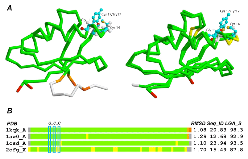
| Figure 4: Structural superposition of the model for protein 11238_88 and coppertranslocating P-type ATPases (CopA). (A.) Left, structural comparison of the model with CopA from Bacillus subtilis, PDB: 1kqk; 1st bar in (B). Right, structural comparison of the model with MerP from Cupriavidus metallidurans, PDB: 1osd; 3rd bar in (B). Positions of the key amino acids: G, C, and Y are shown in cyan rectangles as observed in PDB templates and the model constructed. (B.) Bar representation of structural deviations between four CopA proteins using created model of 11238_88 as a frame of reference. The location of the GxxCxxC motif is highlighted in cyan. The colors of the bars indicate the distance deviation between superimposed corresponding residues using the following color scheme: deviation <2Å, green; <4Å, yellow; <6Å, orange; <8Å, brown; >8Å or not aligned, red; not aligned and terminal residues not aligned, grey. |