 |
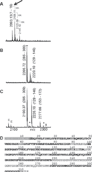
| Figure 5: MALDI ToF MS peptide mapping analysis and amino acid sequence of RA33. [A] Mass spectrum range m/z 2050–2350 shows RA33 epitope peptide in early eluting fraction (fraction 4) upon incubation with anti-His antibody. [B] Mass spectrum of RA33 peptides in late eluting fraction (fraction 8) upon incubation with anti-His antibody. [C] Mass spectrum of tryptic RA33 peptides. [D] Amino acid sequence in single letter code of RA33 showing sequence coverage after trypsin digest in bold letters. The shaded sequence displays the epitope motif that is recognized by the ant-His antibody. Matrix: DHB, E: derived from trypsin autoproteolysis, •: sodium adduct, Δ: potassium adduct, *: oxidation. |