|
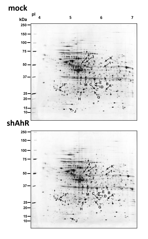
| Figure 2: Representative 2DE gel maps for mock and shAhR cells. A total of 100 µg of proteins were separated by 2DE using the first dimension of 18 cm pH 4-7 linear strips and the second dimension of 8-16% gradient SDS-PAGE. Separated proteins were visualized by silver staining. The spots were analyzed with ImageMaster software and identified by MS/MS sequencing. Down-regulated and up-regulated proteins are labeled as Arabic numerals and English alphabet, respectively. |