|
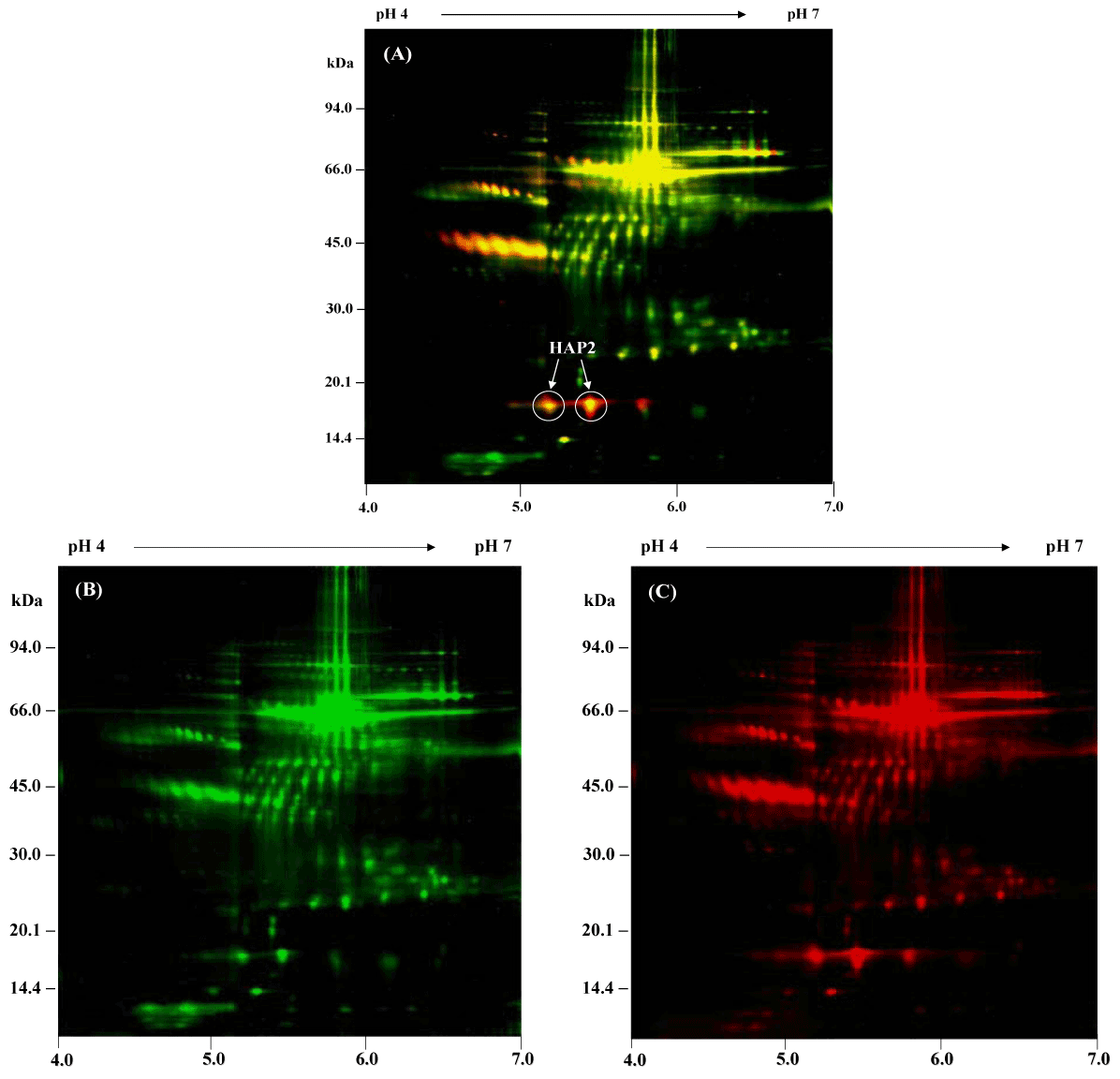
| Figure 3: 2-D DIGE of serum proteins from normal and NSCLC serum samples. Serum proteins from normal serum were labeled with Cy3 (green) and serum proteins from NSCLC serum were labeled with Cy5 (red). Both samples were separated on 2-DE using a narrow range of IPG strip pH 4-7. (A) 2-D DIGE overlay image of Cy3- and Cy5-labeled proteins in normal and NSCLC serum samples. Spots are an equal intensity between the two channels appeared yellow in the overlay image. (B) The normal serum proteins were labeled with Cy3 (green color). (C) The proteins in lung cancer were labeled with Cy5 (red color). The arrows indicate the differential expression of HAP2 isoforms. |