|
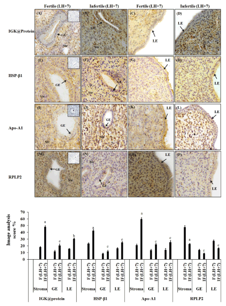
| Figure 4: Representative images (upper panel) showing immunohistochemical localization of IGK@ protein, HSPβ-1, Apo-A1and RPLP2 proteins in fertile (LH+7) and infertile (LH+7) human endometrium. For negative controls (see inset in A, E, I, M) mouse-IgG/ rabbit IgG was used in place of primary antibody. Lower panel shows image analysis of IGK@protein, HSPβ-1, Apo-A1, and RPLP2 in fertile and infertile endometrium. Staining intensity of all these proteins were quantified by image analysis software Image-Pro Plus 4.0 (Maryland, USA). Str (Stroma), GE (glandular epithilium), and LE (luminal epithelium). Magnification X 400, bar=25 μm, n=5 in all the groups. |