|
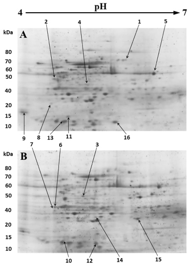
| Figure 1: Representative images of 2-DE gels of WT and Fabp7 KO astrocytes stained with fluorescent dye (Flamingo Gel Stain). Protein spots whose expression levels were different between Fabp7 KO and WT astrocytes are numbered and pointed out by arrows. Nine protein spots show reduction and seven other protein spots represent higher expression in Fabp7 KO astrocytes (B) in comparison with WT counterparts (A). Spot number 16 expected to be FABP7 due to its absence in the Fabp7 KO gel. |