|
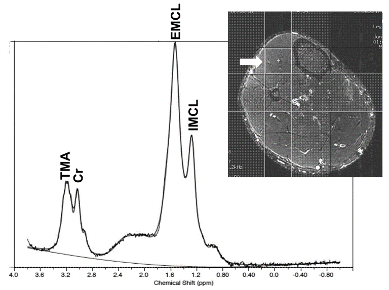
| Figure 1: Sample suppressed 1H spectrum from a healthy skeletal muscle region of the anterior tibialis. Intra-myocellular (IMCL) and extra-myocellular (EMCL) lipid signals, trimethyl ammonium (TMA) and creatine (Cr) peaks are evident. Upper insert shows the axial calf slice with the location of the voxel (arrow) that was used to collect the spectrum. |