|
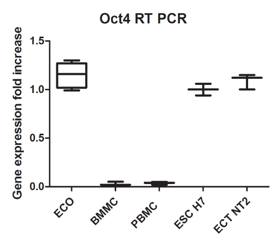
| Figure 10: Quantitative analysis of the OCT4A expression in the ECO cells. The RNA from the cells was reverse transcribed into cDNA, which was then amplified by RT qPCR with the primers for the OCT4A. The amplification was calculated in relation to GAPDH and compared relative to the ESC expression. Labels: ECO – patients’ samples (n=6); PBMC – peripheral blood mononuclear cells (n=6); BMMC – bone marrow mononuclear cells (n=6); ESC – cultures of the human embryonic stem cells H1, H7, H9 (n=3); ECT -cultures of the human testicular embryonal carcinoma cells NT2D1 (n=1). Statistical significance was accepted at the p < 0.001. The samples were run in triplicates. |