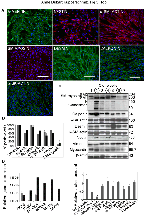
A ) Immunofluorescence studies for the representative clone c4FL3 using antibodies against vimentin, nestin, α-smooth muscle actin (α-SM-actin), SM-myosin, desmin, calponin and α-sarcomeric actin (α-SK-actin). Bars: 25µm.
B) Percentage of positive cells for the different markers in immunofluorescence studies like those illustrated in A. Values are expressed as mean ± SEM. Results for primary layers (n=9, gray bars) and clones (n=7, black bars). Differences are not significant.
C) Western blot analyses. Upper panel: Western blot analyses for SM-myosin, caldesmon, calponin, α-SK actin, desmin, α-SMA, nestin, vimentin and myocardin in 7 clones. Encircled numbers indicate clones c4FL3, c3FL8 and c7FL8. Lower panel: corresponding densitometry data (mean ± SEM; value for each protein related to that of β-actin).
D) Q-RT-PCR data for transcription factors implicated in skeletal muscle differentiation (PAX3, PAX7 MYOD1, MYOG, MYF5, MYF6) in 3 clones (c4FL3, c3FL8 and c7FL8) and in normal human cultured myoblasts. Values are expressed as mean ± SEM of gene expression normalized to that of GAPDH (ΔCt method). Gray bars: clones; Black bars: myoblasts.