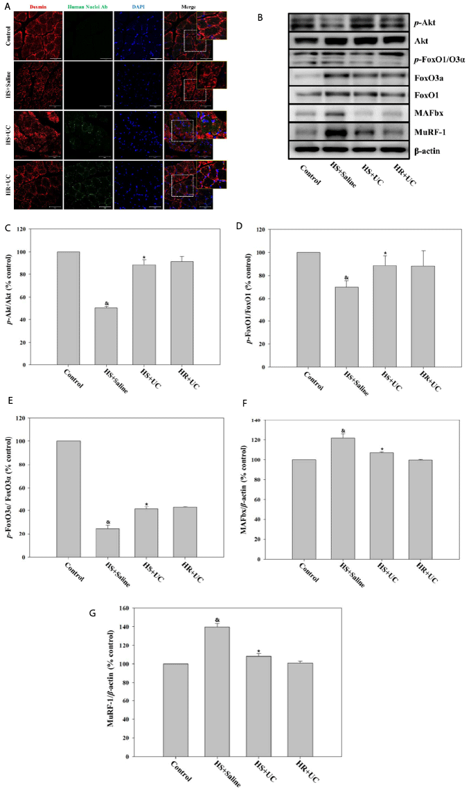
(A) Immunofluorescence analysis using paraffin sections of soleus muscles from the Control, HS+Saline, HS+UC and HR+UC group rats at 2 weeks. Desmin is labeled with Cy3 (red), UC-MSCs are labeled with alexa 488 (green), and nuclei are stained with DAPI (blue). (Original magnification 200X, scale bars=50 μm). (B-G) Immunoblot analysis of the expression of PI3K/AKT signaling pathway. Representative Western blots showing the expression of whole protein level and phosphorylated (p) forms of AKT and FoxO-1/3a, and the expression of total form of MAFbx (Atrogin-1) and MuRF-1 in the soleus muscle. The expression of phosphorylated AKT (C), FoxO-1/3a (D and E), MAFbx (F) and MuRF-1 (G) normalized to β-actin expression (&P<0.05, compared to control group; *P<0.05, compared to HS+Saline group). Error bars represent standard deviation.