|
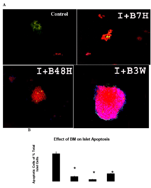
| Figure 4: Islets cultured with BM for 7 hours, 48 hours, and 3 weeks and islet only culture for 7 hours as control were measured by TUNEL assay (green) for apoptosis following pro-insulin fluorescent (red) immunohistochemistry to identify islet β cells. The images on the top left indicate that islets co-cultured with BM (bottom row) have fewer apoptotic cells and stronger insulin staining than those in islet-only culture (top row) (I: Islets Only; I+B: Islet Co-cultured with BM; xH: Culture Hours; xW: Culture Weeks). b. Quantification of apoptotic cells as a percentage of total islet β cells in co-culture groups and human islet culture only groups for 7 hours, 48 hours and 3 weeks. *p<0.01vs. islet only culture (I7 h and I3 W). |