|
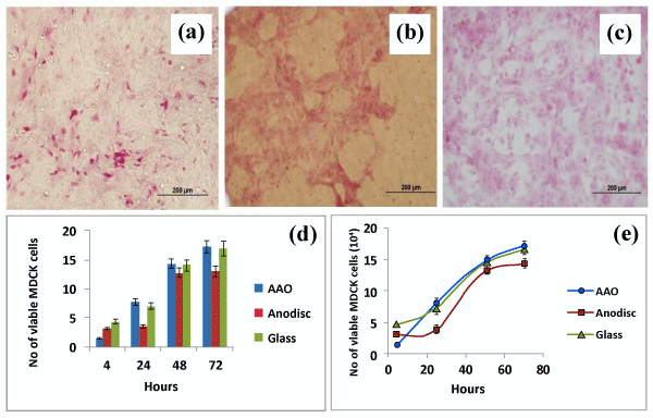
| Figure 2: Optical microscopy of cells proliferation at the 24 h period; a) inhouse AAO membrane; b) Whatman® anodisc membrane; c) glass substrate control; d) a typical 72 h cell proliferation assay; and e) comparison between the number of viable cells on an in-house AAO membrane, Whatman® anodisc membrane and a glass control. Scale bars in (a), (b) and (c) are 200 µm. |