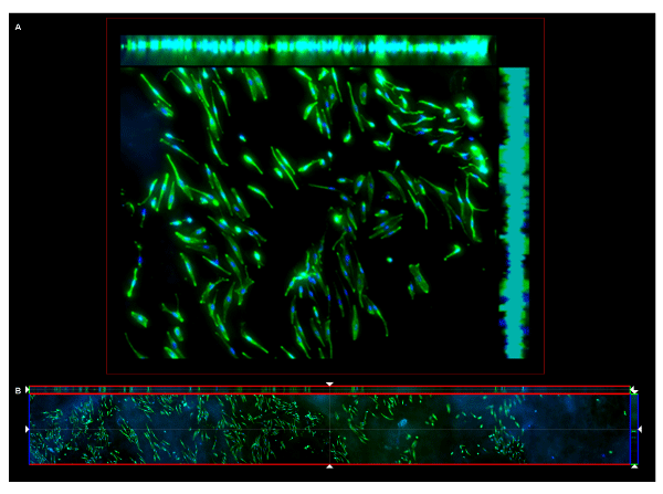
 (A) Maximum Intensity Projection (MPI) z-stacked take of HM on CCC
membrane. Side projections display the depth of the scaffold reconstructed
from 10 consecutive images taken with a 10 μm progression interval. The
cells populated the 100 μm depth of the CCC scaffold. Over 97% of the
cells expressed, the gp100 variant NKI-beteb, present in melanosomes of
ORS melanocytes (HM). (B) ‘cross-census’ panoramic image of 7 images
consecutively taken along the same axis across the full width of a mounted
slide.
(A) Maximum Intensity Projection (MPI) z-stacked take of HM on CCC
membrane. Side projections display the depth of the scaffold reconstructed
from 10 consecutive images taken with a 10 μm progression interval. The
cells populated the 100 μm depth of the CCC scaffold. Over 97% of the
cells expressed, the gp100 variant NKI-beteb, present in melanosomes of
ORS melanocytes (HM). (B) ‘cross-census’ panoramic image of 7 images
consecutively taken along the same axis across the full width of a mounted
slide. |