|
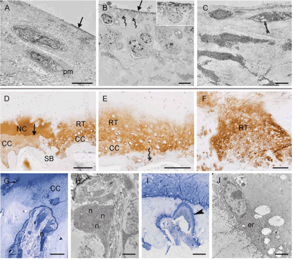
| Figure 7: Upper and lower boundary of the RT. A-C: TEM images of the surface of the RT in treated defect (A, B) and the control (C). A: Elongated chondrocytes surrounded by a pericellular matrix (pm) are arranged parallel to the joint surface. The cells are surrounded by dense ECM, which is covered by a thin layer of electron-dense matrix (arrow). B: The cells at the surface extend cell extensions (wavy arrows and insert) and lack a pericellular matrix but are also embedded in dense matrix covered by dark material (arrow). C: Polygonal cells with many cell extensions and extended ER (arrow) are embedded in fibrous matrix. No electron dense material is visible at the surface. D-F: Immunohistochemical reactions against collagen type II in the deep region of transplanted defect show local differences of RT, CC and SB. D: Residual, homogeneous and intensively stained NC above a homogeneous, brighter-staining CC and unstained SB. The arrow indicates the tide mark. Adjacent to this there is collagen type II positive but somewhat fibrous RT. The underlying CC is hardly distinguishable from the RT but slightly brighter. A similar situation is also visible at the left side in E. E: In the middle of the image the trabeculae of the bone are very thin and the partially decalcified cartilage extends down to the bone lacuna of the subchondral bone (wavy arrow). F: Strong staining of the RT adjacent to a perforation of the SB. G,I: Toluidine blue and H,J: TEM images of cellular activity in the SB in transplanted defects. G,H: Osteoclasts with several Nuclei (n) are resorbing the SB such that the bone marrow lacuna penetrates the CC. I, J: Osteoblasts with abundant ER (er) deposit osteoid (arrowhead). Images A, B, D, E, I and J were taken form cartilage defect treated with a double sandwich-like layer of a hyaluronan web and a collagen type I/III fleece, C from the control defect without transplant, F from the defect treated with a single collagen type I/III fleece layer and G, H with chondrocytes on a single hyaluronan web layer. Scale bars: (A-C) 5 µm, (D-F) 100 µm, (G) 30 µm, (H) 10 µm, (I) 50 µm, (J) 2 µm. |