 |
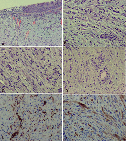
| Figure 2: The lesion is composed of spindle cells with myxoid background (A).The spindle and elongated nuclei with eosinophilic cytoplasm (B and C) are noted with prominent lymphoplasmacytic infiltrate (B and D). The spindle cell components are positive for SMA (E) and Alk-1 (F). |