Tian Meng and Menghua Dai
DOI: 10.4172/2165-7920.1000827
Background: The etiology and pathogenesis of hyperlipidemic pancreatitis are not yet fully understood, and the relationship between the severity and different causes of this disease is still not clear. Now the treatment of hyperlipidemic pancreatitis is mainly to reduce the elevated serum triglyceride and control the secondary risk factors. For severe hyperlipidemic pancreatitis, which can deteriorate in a very fast speed, more efficient and effective therapeutic methods are needed. Case presentation: The patient was a 32-year-old Chinese woman in her 32-week gestation. She was previously healthy, and came to have a sudden onset of acute pancreatitis induced by hyperlipidemia. Her situation deteriorated rapidly, and she simultaneously developed fetal death, respiratory failure, acute renal injury, and severe infections in the lungs, abdominal cavity and the wound. Besides traditional therapies, we used plasmapheresis treatment and successfully saved her life. Conclusion: In recent years, hyperlipidemic pancreatitis and hyperlipidemic pancreatitis during pregnancy seem to happen more frequently than before. For the extremely severe hyperlipidemic pancreatitis, plasmapheresis seems to be a safe and efficient therapeutic approach, and is often needed to save the patients’ lives. However, evidence for evaluating the efficiency and safety of plasmapheresis in the treatment of hyperlipidemic pancreatitis is still scarce, and we think high-quality, large-scale RCTs in this field are needed for further studies. In addition, by reporting this case, we want to remind general readers to pay attention to adopting a healthy lifestyle, and to encourage researchers and healthcare professionals to continue this research.
Grob K, Fretz Ch, Kuster MS, Gilbey H and Ackland T
DOI: 10.4172/2165-7920.1000828
Introduction: Muscle strains within the quadriceps muscle group are common and occur mostly in the rectus femoris. We report a case of an isolated rupture of the tensor vastus intermedius (TVI); a muscle that has recently been described. It belongs to the quadriceps muscle group and is closely related to the vastus lateralis and vastus intermedius. Case presentation: A healthy 62-year old woman presented with a history of a sudden onset of left knee and thigh pain after stumbling and preventing a near fall. Rupture of the aponeurotic tendon of the TVI was diagnosed by magnetic resonance imaging (MRI). Conservative treatment was successful. Four months after injury the patient returned to her pre-injury level of activity. MRI evaluation five months post-injury revealed full resorption of the muscular haematoma and a healed TVI aponeurosis with scar tissue formation. Conclusion: Due to its anatomic appearance, lesions to the TVI may be overlooked. The isolated rupture of the TVI in the present case further supports the recent finding, that the TVI is a distinct anatomical structure independent of the adjacent vasti.
DOI: 10.4172/2165-7920.1000829
Objective: To present a case series of sciatic pain in young doctors, produced by external compression of the sciatic nerve by a wallet.
Summary of background data: Sciatic pain is a common problem in adults and it may be caused by various intraspinal or extraspinal pathologies. Disc rupture and degenerative spine disease are the most common intraspinal causes, while extraspinal etiology remains very rare.
Methods: Description of two cases with sciatic pain produced by external compression of the sciatic nerve at the gluteal region is presented, with review of the relevant literature.
Results: Complete resolution of the sciatic symptoms was achieved at 3 months following removal of the wallet from the back pocket.
Conclusions: When sciatica is the only clinical finding, especially in the young patient, extraspinal causes should be suspected, as early diagnosis could reduce suffering and discomfort of the patient and prevent unnecessary procedures and investigations.
Naveed Ali, Salman Farhat, Syed Imran, Mustafa Jafri, Aamir Ahmed and Faizan Malik
DOI: 10.4172/2165-7920.1000830
An 86-year-old male with a history of metastatic castrate sensitive prostate cancer received a single dose of Denosumab for his bony metastases. Two weeks later, he presented to the hospital due to left foot cellulitis and was incidentally found to have profound hypocalcemia whereas his serum calcium was normal at the time of Denosumab injection. A thorough workup was undertaken which showed severe Vitamin D deficiency. He was diagnosed with Denosumab induced hypocalcemia with underlying Vitamin D deficiency which was refractory to supplemental calcium and Vitamin D. This case demonstrates the potential of Denosumab to cause profound hypocalcemia which can be resistant to therapy. Bone metastasis is a common clinical encounter and Denosumab is an effective therapy to prevent skeletal related events (SRE). Therefore, given its widespread use, it is extremely important to identify and treat risk factors that may aggravate hypocalcemia when treated with Denosumab.
Kassab P, Nara F, Ilias EJ, Castro OAP, Thuler FR, Freitas W, Mancini FC, da Silva PF and Malheiros CAM
DOI: 10.4172/2165-7920.1000831
The possibility of preserving the duodenum and pancreas in the uncinate process neoplasms is, in general, reserved to less aggressive tumors. The literature describes this type of surgery for intraductal papillary mucinous neoplasms (IPMNs), or for cystic tumors.
Bayan Hafiz, Ashwaq Al-qurani, Abdulelah Al-Malki, Esraa Kheel and Ghada Esheba
DOI: 10.4172/2165-7920.1000832
Hydatid disease is caused by genus Echinococcus, it's transmitted to human through sheep and cattle. People who lived in an endemic area should be suspected to have the disease. Pulmonary hydatid disease can be presented by respiratory manifestations as in our case. We report a case of a child, 13 years old, who was presented by shortness of breath and non-productive cough 2 months ago. The patient had an attack of hemoptysis 3 months ago but there is no history of fever, other constitutional symptoms or any medical illness. The patient had a close contact with horse. On examination, the patient was oriented and vitally stable. Both sides of the chest were moving equally with decreased air entry on the left side of the chest. Cervical lymph node enlargement was also detected. The case was provisionally diagnosed as tuberculosis. The x-ray was normal, while CT scan showed two cysts on the left side. The patient was treated surgically with resection of both cysts without lobectomy. Bronchoalveolar lavage was done and together with plural effusion and both cysts were sent for histopathology. The patient received the following medication: Albendazole 200 MG/BID/Orally for 30 days and Cefuroxime 250 MG/Q12H/ Orally for 10 days.
Jing Loong Moses Loh, Zubin J Daruwalla and Mark Chong
DOI: 10.4172/2165-7920.1000834
We report the case of a 14 year old girl admitted with a two day history of left sided thigh and buttock pain and fever. There was no preceding history of trauma. Physical examination revealed a temperature of 38.7°C, tenderness over the left sacroiliac joint, pain on manipulation of the joint and limited abduction and external rotation of the left hip. Radiographs of the sacroiliac joints were normal while an MRI pelvis showed signs consistent with a diagnosis of left sacroiliitis. Although initial blood cultures were negative, intravenous ceftriaxone and cloxacillin were commenced. Symptoms persisted with subsequent MRI showing an adjacent abscess. The CT guided aspiration of the abscess with a study of synovial fluid demonstrated the presence of Salmonella (non-typhi species). A second set of blood cultures revealed the same pathogen and confirmed sensitivity to ceftriaxone. Antibiotics were tailored to this result. She received intravenous ceftriaxone for a total of 90 days. On completion of intravenous antibiotic therapy, there was significant resolution of pain and improvement in ambulation. MRI findings were consistent with resolution of infection. Oral cefixime was continued for a total of 30 days. Her condition resolved with no recurrence of symptoms at the time of last follow up.
Rifki Albana, Jelita A Purba, Heriyanto Manihuruk and Mohd Ariff
DOI: 10.4172/2165-7920.1000835
Most tuberculosis infections of the musculoskeletal system occur in the spine, hip or knee with Hip tuberculosis (TB) constituting 15%-20%. Highest rates of the tuberculosis cases in Indonesia occur in Papua. Neglected cases or delayed treatment can make management challenging. We present a 47 years old Papuan man who presented with progressive destructive TB of the hip due to lack of resources and access to adequate healthcare. He underwent a limited duration of anti-tuberculosis regimen and subsequently had debridement, girdlestone, and cemented total hip arthroplasty (THA) of the affected hip. From this case we conclude that hip arthroplasty in tuberculous arthritis of the hip with compliant administration of anti-tuberculosis drugs is a safe procedure with either cemented or cementless implants.
DOI: 10.4172/2165-7920.1000836
We reported strangulated male external genitalia by three steel rings for 24 hours. It is difficult to manage without adequate device and some rescue devices may bring possible iatrogenic injury to genital organ. Steel ring was removed by hydraulic cutter instead of angle grinder which was applied in our previous experience and mentioned by literature. Angle grinder may bring cutting or thermal injury to patient or medical staff. It may be safer method to rescue strangulation with hydraulic cutter instead of angle grinder especially in those patients with blood borne disease. However, management may be individualized for difference material and shape of constricting device.
Luis Balmore Gutierrez, Tara A Morgan, Thomas Link, Brian Feeley and Daria Motamedi
DOI: 10.4172/2165-7920.1000837
We report a case of suprapatellar fat pad impingement diagnosed and treated using musculoskeletal ultrasound. This report describes the utility of musculoskeletal ultrasound in making the diagnosis of suprapatellar fat pad impingement, which may potentially present as anterior knee pain, and more specifically, anterior superior knee pain. The importance of recognizing this entity on musculoskeletal ultrasound, especially when the imaging findings are subtle on prior MR imaging, is emphasized. Unconvincing suprapatellar fat pad edema on MR imaging, for example, should not preclude consideration of this entity at targeted ultrasound of the anterior knee in a patient with anterior knee pain. Furthermore, once the diagnosis is made using ultrasound, the suprapatellar fat pad impingement can be immediately and effectively treated with ultrasound-guided injection of steroid and anesthetic.
Kevin Landefeld M.D, Holly Gonzales M.D. and Gary E Sander M.D. Ph.D.
DOI: 10.4172/2165-7920.1000838
62 year-old female presents with hypertensive urgency while taking daily NSAIDs. This case demonstrates the effect of NSAIDs on BP, an often over-looked etiology of secondary hypertension. The detrimental effects of NSAIDs upon blood pressure have been well documented. The report reiterates and reviews the severity of the problem. We will review the existing literature and discuss the importance of small increases in blood pressure.
Nimat Alam, Linda Esteban, Enrique Tobias and Jenet George
DOI: 10.4172/2165-7920.1000839
Thyrotoxic periodic paralysis (TPP) is characterized by abrupt onset of hypokalemia and paralysis. This condition primarily affects the lower extremities but may affect both upper and lower extremities and is secondary to thyrotoxicosis. It is most commonly seen in Asian men and it has been increasingly reported in USA due to the rise in the immigrant population. Hypokalemia in TPP results from an intracellular shift of potassium induced by the thyroid hormone sensitization of Na+/K+ATPase rather than depletion of total body potassium.
Javeed Iqbal, Ikhlas Ahmad, Md Asif Ahmed and Ambreen A Ahangar
DOI: 10.4172/2165-7920.1000840
Fanconi-Bickel syndrome (FBS) is a rare inherited glycogen storage disease (GSD) caused by defects in facilitative Glucose Transporter (GLUT2) gene that codes for the glucose transporter protein 2 expressed in hepatocytes, pancreatic beta cells, enterocytes, and renal tubular cells. The clinical picture is characterized by glycogen accumulation in liver and kidney resulting in hepatomegaly and nephromegaly, impaired utilization of glucose and galactose, proximal renal tubular acidosis, hypophosphatemia rickets, and short stature. This is an autosomal recessive disorder discovered in 1949 and the pathogenic mutation of GLUT 2 gene of hepatocytes, beta cells of pancreas and renal tubules were discovered in 1997.
Xiuqing Zhang, Bindong Song, Guixia Xu, Shengtang Zhang, Yinyong Ding and Fangli Cao
DOI: 10.4172/2165-7920.1000841
Background: Cerebral Salt Wasting Syndrome (CSWS) refers to the process of intracranial lesions, sodium salt loss by hypothalamus-renal pathway and caused clinical manifestations syndrome of high urinary sodium, hyponatremia, hypovolemia. The clinical manifestations and laboratory are similar to syndrome of inappropriate antidiuretic hormone secretion (SIADH). It is easy to be misdiagnosed.
Case description and management: In this paper, the clinical data of one case of cerebral salt wasting syndrome was diagnosed. By given fluid and sodium replacement, the condition can improve quickly. Through analyzing and reviewing related literature, the understand of diagnosis and treatment of cerebral salt wasting syndrome can be improved.
Conclusions: The main clinical manifestations of cerebral salt wasting syndrome are hyponatremia, high urine sodium, diuresis, hypovolemia. Cerebral salt wasting syndrome in children has very high risks and we should improve the understanding. Early diagnosis and prompt treatment are very important.
Yadav S, Deane AKS, Hasan S and Hayer P
DOI: 10.4172/2165-7920.1000842
Milwaukee shoulder syndrome is a progressively destructive shoulder arthropathy mostly seen in mostly women over 70 years age. It is associated with severe pain and generalized restriction of joint movements, usually associated with rotator cuff tear. A 85 years old female presented to our hospital with chief complain of pain and decreased range of movements in both shoulder joints. Investigations were done including FNAC and Biopsy which could not identify any evident cause. Therefore diagnosis was made on clinico-radiological bases and patient was managed. Surprisingly patient had relief with plain NSAID’s compared to opioids.
Vandita K Patil, Usha Varghese, Kailas N Patil, Swayed Mahmud Ali Reza, Jawaher Al Yazeedi, Shaji Varghese and Ashwin Varghese
DOI: 10.4172/2165-7920.1000843
A 27 years old healthy female of twenty eight weeks pregnancy with history of low grade fever and dry cough for one day presented with intrauterine fetal death. Following spontaneous preterm delivery of the dead fetus, within three hours, patient developed irritable cough, dyspnea, tachypnea, restlessness and cyanosis. She was put on face mask with oxygen flow of ten litres/minute and was nebulized with Salbutamol in the delivery suite but gradually she developed desaturation of 76%. As the condition of patient was worsening patient was transferred to intensive care unit. In intensive care unit patient was intubated and put on ventilator immediately. X-ray chest was showing bilateral infiltrates and arterial blood gases was showing PaO2/FiO2 ratio of 55.6%. Pulmonary capillary wedge pressure was not checked as pulmonary catheterization is not practiced in our Intensive care unit. Cardiogenic component of pulmonary edema was ruled out indirectly by history, electrocardiogram, echocardiography, central venous pressure and X-ray chest (heart shadow). She was diagnosed as a case of severe acute respiratory distress syndrome (American European consensus conference guideline, 2012) and non-cardiogenic pulmonary edema due to amniotic fluid embolism syndrome. In course of management, maximum emphasis was given on lung protective ventilation and fluid, conservative strategy along with medical and other supportive management. On her eighth day on ventilator, she was extubated and on ninth day, she was shifted to maternity ward. On fourteenth day she was discharged from hospital. She came for follow up after one month of her discharge and was found to have no residual complication.
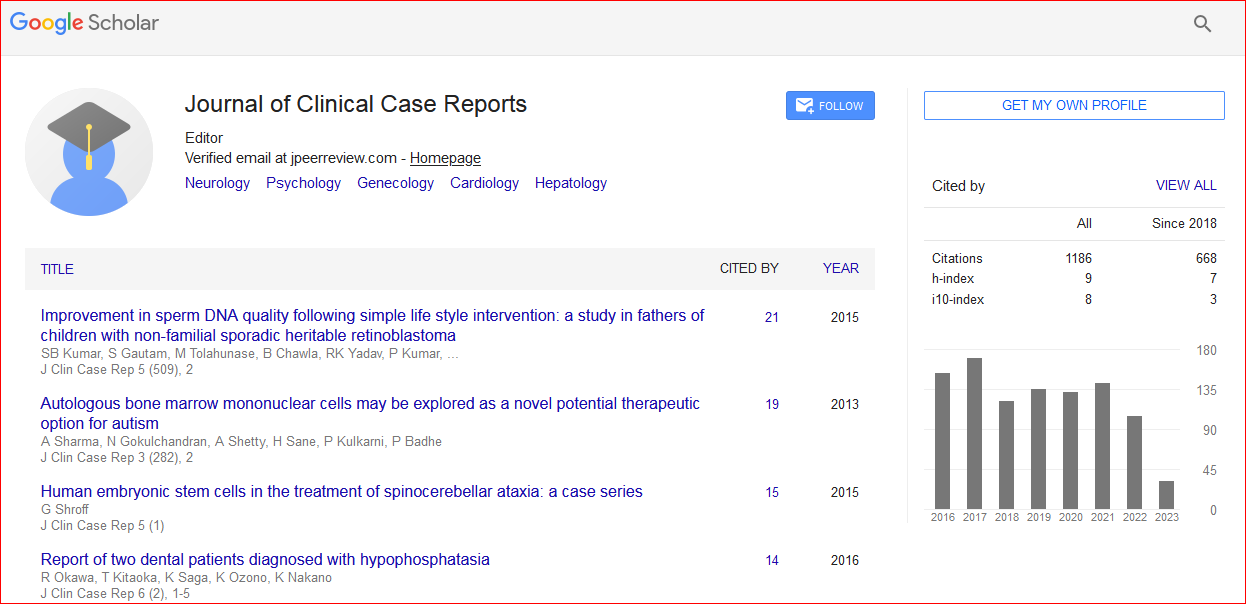
Journal of Clinical Case Reports received 1295 citations as per Google Scholar report

 Spanish
  Chinese
  Russian
  German
  French
  Japanese
  Portuguese
  Hindi
Hindi 