Suresh Raghavaiah, Hardev Ramandeep Singh Girn, Parshotam Lal Gautam and Siddarth Prakash
DOI: 10.4172/2167-1222.1000252
Most liver injuries after blunt trauma abdomen are currently treated by non-operative management (NOPM)1-3. Delayed haemorrhage necessitating operative intervention is one of the main complication following NOPM3. We report the case of a young male who suffered delayed fatal exsanguination after NOPM of a high grade liver injury and review the available literature regarding management of high grade liver trauma.
Mohamad Gouse, Subin Babu, Ravichand Ismavel and Korulla Mani Jacob
DOI: 10.4172/2167-1222.1000253
A segmental bone defect caused by an open fracture is a daunting problem to deal with and the traditional treatment options that are currently practiced to treat such conditions include allo or autologous bone graft, distraction osteogenesis and membrane induction. Infrequently the course of treatment is complicated by non-union and infection, thus requiring multiple staged procedures that may affect the functional outcome. We present a case of a 25 year old gentleman who had sustained an isolated open type III A comminuted distal femur fracture with a part of the diaphyseal distal femur extruding from the body with no soft tissue attached. His plain radiograph showed comminuted distal femur AO C3 fracture. Following debridement, the extruding diaphyseal fragment was washed and cleaned with normal saline and retained inside the body. The limb was stabilized with an external fixator and 48 hours later he was taken for a second debridement wherein a stable internal fixation was done retaining the extruded fragment. Post operatively he recovered well without wound complications or any deep or superficial infection. The follow up radiographs showed that the bone coalesced with an uneventful union and in next 3 years the knee ROM was 0-130 degrees. His lower extremity functional score was (LEFS) 74. This case is to highlight the importance of judicious insight at times when debridement is the matter of concern, taking into consideration that all reconstructive options are available. The outcome of other staged reconstructive procedures to bridge such bone loss is far more tedious and sure to have a far less efficacious outcome than what has been described here. This report also described about other literature about the option available for such condition.
Purushothama Sastry, Sujana Theja JS, Supreeth N and Arjun Markanday
DOI: 10.4172/2167-1222.1000254
Introduction: Called Lover’s Fractures, the calcaneus commonly fractures due to fall from height. The calcaneus is the most frequently fractured tarsal bone. Tarsal bone fractures account for about 2% of all adult fractures. Of these, 60% are calcaneus fractures.The heel bone is often injured in a high-energy collision where other parts of the skeleton are also injured. In up to 10% of cases, the patient can also sustain a fracture of the spine, hip, or the other calcaneus. Injuries to the calcaneus often damage the subtalar joint and cause the joint to become stiff. This makes it difficult to walk on uneven ground or slanted surfaces. Case Report: A 24 year old male, working as a lift operator presented to the casualty of the hospital after the elevator broke down and came down in a free fall from about a height of 4 stories (35 feet). He presented along with the other occupants of the lift who also sustained calcaneal fractures.The case on arrival was subjected to ATLS protocol and through radiologic work up was done. Following a period of 12 days post trauma he was operated for bilateral calcaneal fractures and discharged 10 days post operatively. Conclusion: Calcaneal fractures continue to tread the fine line between operative and conservative management. In a young individual an operative management is likely to give a better outcome as the associated soft tissue problems, commonly encountered, yield a better outcome and faster healing rates.Used of closed reduction further helps alleviate the need to worry about wound healing
Umesh Kumar Meena, Anil Kumar Sharma, Prateek Behera, Piyush Kumar Kanani and Devendra Singh
DOI: 10.4172/2167-1222.1000255
Army recruits are prone to injuries of the musculoskeletal system primarily due to the sudden increase in physical activities. While stress fractures of the metatarsals, tibia and other lower limb bones as well as injury of tendons is common, triceps tendon avulsion in an army recruit has not been reported in literature. As such, triceps tendon avulsion is an uncommon injury. We are reporting a case of a 24-year-old army recruit who had an avulsion of triceps tendon during training and which was missed initially. We managed him surgically by using non-absorbable Ethibond sutures. The patient recovered his full range of elbow motion and at the end of one year had no functional limitations. A physician needs to have high degree of suspicion to avoid delayed diagnosis and prolonged disability. Surgical repair of complete ruptures using transosseous sutures leads to excellent functional results and would probably be the best method of managing these injuries.
Shahid Hussain, Hamid Rather and Asif Qayoom
DOI: 10.4172/2167-1222.1000256
Objective: The clinical experience with single intra-articular hyaluronic acid injection in knee OA is limited. The aim was to assess the therapeutic efficacy, tolerability and adverse events of single-shot intra-articular hyaluronic acid injection in knee OA.
Method: Between February 2008 and December 2010, forty eight (48) patients (Males=21, Females=27) with OA knees were enrolled in this prospective study. These patients had failed to respond adequately to conservative treatment including analgesics and rehabilitative modalities. The inclusion criteria were: 1) Resting visual analog scale pain of >50 mm and 2) Radiographic evidence for osteoarthritis and at least one of the following three characteristics; Age ≥50 yrs, Morning stiffness ≤ 30 min in duration and coarse crepitus on motion (as per American College of Rheumatology guidelines 1986). Functional scoring as per WOMAC and VAS for pain at rest and during walking was documented. Patient satisfaction was documented as per Linkert scale. Objective analysis included measurement of knee flexion-extension, circumference at the level of suprapatellar pouch and change in joint space width (Tibiofemoral joint). Patients received a single injection of Synvisc-One (consisting of 6 ml hylan G-F 20). The patients were reviewed at 1 month, 3 months and 6 months post-injection with final analysis at 1 year. Use of only paracetamol, when required was allowed for concomitant analgesia but disallowed around the time of clinical evaluation.
Results: The mean age of the patients was 65 ± 5 years; mean duration of symptoms 5.4 ± 1.5 years and mean body mass index (BMI) 29.1 kg/m2. Bilateral knees were involved in 69% of patients. Knee swelling and effusion was present in 70% and 31.5% patients, respectively. All the patients were available at final follow-up. The resting and walking VAS pain significantly improved from baseline after the injection (45 from 70 and 50 from 82, respectively). There was significant improvement of pain and disability based on the WOMAC scores. Adverse events were recorded and included local pain and swelling, mild redness, and/or effusion in the knee. Based on “Knee Society Score” the overall efficacy was judged as excellent in 55%, satisfactory in 43%, and poor in 2%. The beneficial effects stayed till 6 months but returned to baseline at last follow up at 1 year. Substantial improvement was noted in knee flexionextension and knee swelling (p<0.05) but no change was observed in joint space width.
Conclusions: This study confirmed the therapeutic efficacy and safety of single-shot intra-articular injection of hyaluronic acid for the treatment of osteoarthritis of the knee. The pain relief and functional improvement lasts for up to 6 months. The procedure is tolerated well and associated with very few local adverse events. The need for concomitant analgesia is reduced.
Asaf Acker, Amir korngreen, Bloom Shlomo, Shelef Ilan, Perry Zvi, Atar Dan and David Lebel
DOI: 10.4172/2167-1222.1000257
Minimal invasive techniques for fracture repair are becoming more prevalent as increased experience is gained by orthopedic trauma surgeons worldwide, with minimal invasive plate osteosynthesis (MIPO) surgery being a popular one. In the current case report we describe a rare complication of fracture management, where anterior tibial artery occlusion occurred as a result of direct pressure by an LCP 4.5 mm plate inserted via MIPO technique. The case involves a 41-year-old man was brought to the emergency trauma room after sustaining a gunshot injury to the Lt tibia. the surgical team preformed a closed reduction and internal fixation with LCP 4.5 mm (Synthes) plate and screws through a lateral incision in a minimal invasive technique. In the recovery room, thirty minutes after the end of the first surgery, the patient developed acute leg ischemia and was rushed back into the operating room. After lateral fasciotomy and exploration, the tibial peroneal tract was found to be completely torn, with no pulse of the tibialis anterior artery, Thrombectomy was performed and a graft used. Even though, the ischemia continued and Selective angiography demonstrated the tibialis anterior pressed between the tibia and the plate with complete obstruction. The plate was immediately removed and the artery was reconstructed using the saphenous vein from the uninjured leg. Even though arterial complications after fracture management are rare, they do happen and one should keep them in mind and be vigilant for them, thus if they do occur, one can attend to them early.
Boris Kovalenko, Mellisa Roskosky, Brett A. Freedman and Michael S. Shuler
DOI: 10.4172/2167-1222.1000258
Background: Concerns have been raised about the ability of Near Infrared Spectroscopy (NIRS) to monitor skeletal tissue regional oxygen saturation (rSO2) in excessive light conditions, as are found under the overhead lights of the operating room. This study seeks to determine whether varying intensities of ambient light exert an influence on NIRS measurements of skeletal tissue rSO2.
Methods: Thirty people were recruited from the staff of a local surgical center to participate in the study. Three separate NIRS devices (Covidien INVOS Cerebral Oximeter 510°C, Nonin EQUANOX Model 7600, and a CASMED MC-2030C Cerebral Oximeter) were used to obtain readings from the anterior compartment of the leg. Illuminance was recorded simultaneously with oximetry data in an operating room with (1) no lights on, (2) room lights, and (3) surgical lamps set to maximal intensity.
Results: No differences were seen in rSO2 values under the different lighting conditions while using the Nonin device. There was a statistically significant difference between rSO2 for lights off versus room lights (-0.933, p=0.0045) as well as for lights off versus operating room (OR) lamps (level 5) (-0.50, p=0.0035) for the INVOS device, although the INVOS device was not always able to produce a reading in the presence of high-intensity OR light. While there was no difference found between lights off and room lights when using the CASMED device, it was unable to display a value in the presence of high-intensity OR lamp light.
Conclusions: The results indicate that the presence of light has an effect on oximetry readings; however, the presence of such an effect is dependent upon the device being used. While other applications of the device, such as cerebral oximetry, may allow for drapes to cover the areas being monitored, monitoring for compartment syndrome of the leg would not be as forgiving. This application would be best served by a device capable of monitoring at all light levels.
Malgoire JY, Lardière-Deguelte S, Piardi T, Amroun K, Renard Y, de Mestier L and Kianmanesh R
Bilhemia, defined as the passage of bile into the bloodstream through a bile duct-venous system fistula, is a rare complication after liver blunt trauma. We report here the case of a 20 year-old patient who presented with a liver blunt trauma complicated with bilhemia in who hemodynamic stability enabled non-surgical management.
Osman Celbis, Mucahit Oruç, Mustafa Dogan, Bedirhan Sezer Öner, Cihan Göktürk, Nusret Ayaz, Çagatay Önal S and Semih Petekkaya
DOI: 10.4172/2167-1222.1000260
Yasmine Kamhieh, Dimitra Leivadiotou, Dimitrios Manoukian and William Williams
DOI: 10.4172/2167-1222.1000261
Introduction: There is little evidence consensus on the optimum timing for surgical fixation of closed ankle fractures. This powered study compared rates of soft tissue complications in patients operated on within 24 hrs, at 24-48 hrs, or beyond 48 hours from injury.
Materials and Methods: In this retrospective study, N=160 adults with closed ankle fractures were categorised by time to surgery (<24 hrs, 24-48 hrs, and >48 hrs), and post-operative complication rates were compared (power 0.85).
Results: Soft tissue complications were higher following surgery at 24-48 hrs (15%) vs. surgery after 48 hrs (3.6%), P 0.051. There was no other significant difference in any complication rates. The length of hospital stay postoperatively did not differ between groups.
Conclusion: Surgery within 24-48 hours from injury resulted in higher rates of minor soft tissue complications. Further research is necessary to confirm these findings, and to quantify soft tissue swelling and its effect on postoperative complications.
Ravi Kumar Gupta, Anubhav Malhotra, Pawan Kumar and Gladson David Masih
DOI: 10.4172/2167-1222.1000262
The problem of fixation of 3 and 4 part displaced proximal humerus fractures was thought to be greatly solved with the evolution of proximal humerus internal locking system (PHILOS). We report a case of migration of screws of PHILOS plate into the shoulder joint, where salvage was done with hemi-arthroplasty. The decision for osteosynthesis in complex 3 and 4 part fracture needs a high index of awareness of the potential complications of PHILOS.
Johnson A, Roskosky M, Freedman B and Shuler MS
DOI: 10.4172/2167-1222.1000263
Near-Infrared Spectroscopy (NIRS) measures to a depth of 2 to 3 cm below the skin, raising concern over the utility of NIRS in the obese patient. The purpose of this prospective study is to investigate the effect of overlying adipose tissue thickness (ATT) on NIRS oxygenation measurements of skeletal muscle. ATT was measured by ultrasound. NIRS sensors were placed over the anterior and superficial posterior compartments of one leg during exercise and the change in regional oxygen saturation was calculated for each compartment. There was a decreasing trend in change of rSO2 from baseline with increasing ATT. Extremely obese patients (BMI >40) had significantly smaller changes in rSO2 from baseline as compared to otherwise similar patients in both the compartments (p<0.01). As ATT increased, the change of the NIRS values from baseline decreased. There was not a specific BMI or ATT determined to be incapable of being monitored.
DOI: 10.4172/2167-1222.1000264
Jaspreet Singh Chhabra, Ravindra Sabnis, Ankush Jairath, Arvind Ganpule and Mahesh Desai
DOI: 10.4172/2167-1222.1000265
Background: Reactive attachment disorder (RAD) is a psychiatric diagnosis usually made in childhood. It is commonly resistant to treatment, including pharmaceuticals and psychotherapy. A significant clinical feature of RAD is disturbance in affect regulation. This feature of affect regulation abnormality is similar to the over reactivity seen in post traumatic stress disorder (PTSD). A new treatment, called stellate ganglion block (SGB), has been shown to be effective in treating the over reactivity associated with PTSD in the adult population. It involves placing a local anesthetic near a cervical sympathetic ganglion. SGB is believed to reduce sympathetic over activity, which may last for months, leading to improvement in affect regulation. The commonality of abnormal affect regulation (in PTSD and RAD) lead the author to use SGB in a pediatric patient with RAD and in a pediatric patient with PTSD.
Methods: Both patients received a stellate ganglion block on the right side at C6 level utilizing fluoroscopic guidance and 0.5% bupivacaine. Both patients had consent provided for performing the SGB from their parents. The responses to SGB were per parents’, teachers’, and children’s reports.
Results: Both patients experienced immediate, significant and durable relief of RAD and PTSD symptoms, respectively. The PTSD patient requested repeat treatment after three months following added trauma. Both patients markedly reduced antidepressant and antipsychotic medications while maintaining their functional improvements.
Conclusion: Selective blockade of the right stellate ganglion at C6 level is a safe and minimally invasive procedure that may provide durable relief from RAD and PTSD symptoms in a pediatric population, allowing the safe discontinuation of psychiatric medications.
Charles T Price, Michael J. Muszynski, Julie A Zielinski and Charles Stewart
DOI: 10.4172/2167-1222.1000267
Background: Motorboat propeller strikes can cause devastating injury due to injury mechanics and complex contamination. The frequency of these injuries has been under-reported. This paper provides a review of propeller injuries admitted to our hospital over a six-year period. Discussion includes injury mechanics, initial management, literature review, accident statistics, and possible prevention measures.
Methods: Charts of all patients admitted to our hospital following boating injury were reviewed. Thirteen patients were identified with motorboat propeller injuries. A review of boating accident statistics during the same period of time was also conducted.
Results: Patients averaged 2.8 surgical procedures during the initial hospitalization. Infections developed in 46% of patients and there were five amputations in three patients. During the period of this study the number of reported propeller or skeg boating accidents increased from 8 per year to 80 per year in Florida.
Conclusions: Motorboat propeller injuries can be extensive due to severe mechanical damage and contamination with uncommon marine organisms. Blood loss and contamination may be underestimated at time of admission. Therapeutic triple antibiotic management and frequent irrigation and debridement are recommended. Multiple surgical procedures and extended hospitalization are often required with lifelong physical impairment as the final outcome. The apparent increase in reported injuries probably reflects improved accuracy of reporting rather than true increase in frequency of these injuries. Safety education and enforcement along with development and implementation of safety devices is recommended.
Arabo Saidou Mohamadou, Atemkeng Tsatedem Faustin, Tsiagadigui Jean Gustave, Ndando Polle Richard and Bayiha Alphonse
DOI: 10.4172/2167-1222.1000268
Introduction: Complex fractures around the knee are really challenging. They are caused by a direct high-energy mechanism and can be either opened or closed. The main objective of this work was to do a preliminary study of these injuries in our setting.
Materials and Methods: This was a prospective and descriptive study carried out from January 2010 to December 2013, in the orthopaedics and traumatology service of the Laquintinie hospital in Douala. Were included, all patients with around knee fractures, classified 33-A3, 33-C, 41-A3 or 41-C according to the Swiss orthopaedic association (OA) classification.
Results: We had 18 male patients. No female. All were victims of road traffic accidents involving at least one motorbike. The average time of occurrence of the accident was 11 pm. Seventeen lesions were opened with a predominance of the types 1 and 2 of Gustillo and Anderson’s classification. AO Type 33-C3 (5 cases) were more frequent on the femur, while AO type 41-A3 (4 cases) dominate the tibia fractures. Seven patients presented floating knees (above and below knee fractures). The most common associated injurie was the patella fracture (2 cases). Twelve patients including 6 floating knees were treated surgically. Condylar blade plate (7 cases) was the most used at the distal femur whereas in the proximal tibia it was the OA plate (6 cases); four cases were treated conservatively and two fractures were complicated by complex vascular injury imposing amputation at the thigh level.
Discussion: Our study showed that these lesions were not rare; they represented around 2.5% of serious injuries observed during highway accidents caused by the two-wheeled vehicles in our milieu. We had complex lesions in terms of anatomoclinical, therapeutic and functional aspects.
Conclusion: Complex fractures around the knee are becoming frequent in our milieu. They are caused by motorbikes and are difficult to treat.
DOI: 10.4172/2167-1222.1000269
Hernando Rafael and Gabriel Polo
DOI: 10.4172/2167-1222.1000270
Background: Since 1986 by the work of Goldsmith, we know that placing omental tissue on the injured spinal cord, we can provoke neurological improvement.
Case report: A 4-year-old girl received a gunshot wound on August 2010., at the upper cervical cord, which was followed immediately by tetraplegia. Lose of respiratory ahttps://www.omicsonline.org/utomatism and she was connected to a fan. Preoperative MRI scans revealed a severe ischemic infarct at C2-C3. On July 2011, she received an omental transplantation. During surgery we found abundant scar tissue on the cervical cord between C2 and C3, small intramedullary cyst in the left side, reduction of blood vessels, and 70 percent of cervical cord hypotrophied. On this residual cervical cord a segment of omentum was placed. Two days after surgery, she began with respiratory automatism and voluntary movement of shoulders and right limbs. She could stand up and to walk with aid of orthopedic devices since 4 months after surgery. At present, 4 years after surgery, she(a 9-year-old girl) present partial control of sphincters and motor improvement by 40 percent. During the postoperative evolution she receive rehabilitation and electrical stimulation in the cervical cord.
Conclusions: These results indicate that ischemic neurons and axons in the traumatized cervical cord can improve if is revascularized with omental tissue.
Maniglio M, Schweizer A and Nagy L
DOI: 10.4172/2167-1222.1000271
We present the results of a retrospective study looking at postoperative outcomes of treatment of scaphoid nonunions following three different techniques: Palmar (nonvascularized) iliac crest, vascularized bone-grafts dorsal and palmar. The purpose is to present the consolidation rates and discover if range of motion, grip strength and pain differ using the different grafts.
Methods: We evaluated 57 cases with a mean follow-up of 6.6 months. 24 patients had nonunion of the proximal scaphoid pole with avascular osteonecrosis. Operative technique were chosen based on the vascularity, location of non-union, previous operations, and pedicle availability. Conventional graft was used in 19 patients, palmar vascularized in 15 and dorsal vascularized graft in 23. We measured and compared clinical and radiological outcomes.
Results: Out of 57 nonunions 46 united. No significant difference in rate of consolidation was found between the 3 treatment groups. Highest percentage of consolidation, 87%, was in patients treated with a palmar vascularized graft, whereas the consolidation rate of dorsal vascularized graft was 78% and of iliac crest graft 79%. Grip strength improved significantly. The most in iliac crest group (from 70% to 91%) and the least in dorsal vascularized graft group (from 81% to 83%). Best range of motion in flexion/-extension was in the iliac-crest group, although not being significantly. Average Mayo score was 82. 18 patients showed an excellent, 17 a good, 13 a satisfactory and 3 a poor result. No significant difference was found between the groups.
Conclusion: Vascularized grafts had a comparable consolidation rate and outcome in Mayo score as the iliac crest group, even though these cases were more problematic due to the presence of avascular necrosis. Iliac crest grafts afforded the best results concerning grip strength and range of motion. The gain in grip strength and range of motion was less with vascularized grafts.
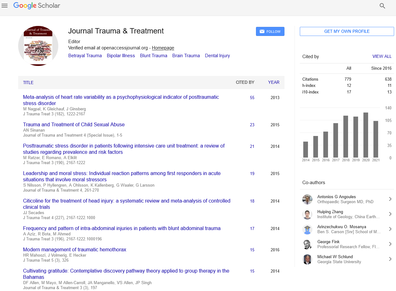
Journal of Trauma & Treatment received 1048 citations as per Google Scholar report

 Spanish
  Chinese
  Russian
  German
  French
  Japanese
  Portuguese
  Hindi
Hindi 