|
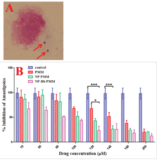
| Figure 3: A) Infection of leishmania to human healthy macrophages and the production of amastigote was established by introducing promastigote to the culture of macrophage at 1:10 ration of macrophage:promastigote. Giemsa stain was used to stain the amastigotes and macrophage nucleus to count the amastigotes under microscope at 100X; where red lines indicated the amastigote cells. B) Graph indicated the percentage inhibition of amstigotes at 48 hrs after treatment of infected macrophages by normal PMM, Cs- Chs PMM and Cs-Chs-Hb PMM; one untreated control was also taken. A significant decrease in LD50 concentration was recorded for Cs-Chs-Hb PMM nanoformulation (75 μM) as compared to Cs-Chs PMM (100 μM) and PMM (130 μM). Samples were analyzed by two ways Anova with statistical differences indicated as follows: p<0.001, *** for control vs. Cs-Chs PMM (140 μM), p<0.001, *** for control vs. Cs-Chs Hb-PMM (120 μM) and p<0.05,* for PMM vs. Cs-Chs Hb-PMM (120 μM). |