|
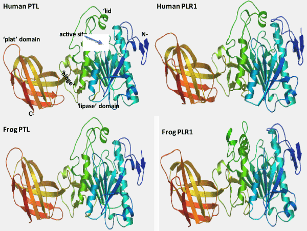
| Figure 4: Tertiary Structures for Human PTL and PLR1 and Frog PTL and PLR1 Sequences. Tertiary structures for human PTL and PLR1 were obtained from Winkler et al. [12] and from a submitted report to the PDB data bank (2PPL) respectively, and using SWISS MODEL methods for the frog PTL and PLR1 sequences; the rainbow color code describes the known tertiary structures from the N- (blue) to C-termini (red color); arrows indicate directions for β-sheets; known or active site for human PTL; N-terminal and C-terminal regions are shown, as are predicted structures and locations for ‘lipase’ and ‘plat’ domains; the ‘lid’ covering the active site; and the ‘hinge’ separating the 2 domains. |