|
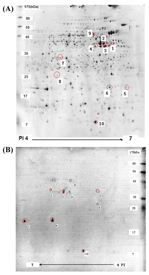
| Figure 2: 2-D Western blot analysis of the total membrane proteome of Chinese Brucella vaccine strain M5. The 2-D gel was electrobloted onto PVDF membrane (20V, 1h) and western blotting analysis was performed with antisera from Brucella-infected goats. A total of ten immunodominant protein dots were recognized by antiserum and labeled with the Arabic numbers on the 2-D gel (Figure 2-A) and PVDF membrane (Figure 2-B), respectively. |