|
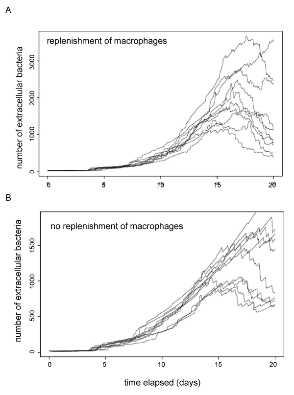
| Figure 5: Comparison to simulation of M. tuberculosis model. Time course of infection is plotted as in figure 3. Initial macrophage number set to 100, dissemination probability set to 5%. Number of extracellular bacteria in the lung (black lines) and liver (red lines) over 20 days with (A) or without (B) replenishment of macrophages. |