 |
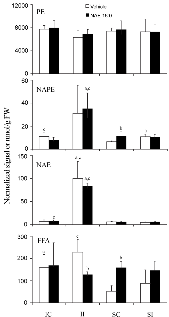
| Figure 2: Ischemia/reperfusion- and sham-operation-induced changes in total PE, NAPE, NAE, and FFA content in contralateral (IC and SC) and ispsilateral (II and SI) brain tissues of rats treated with vehicle or exogenous NAE 16:0. Data are shown as mean ± SD. Statistical significance (P < 0.05; N = 4 (for NAE samples N = 6)) between contra- and ipsilateral tissues is indicated by ‘a’, and vehicle and NAE 16:0 treatment by ‘b’ and ischemia and sham tissues by ‘c’ as determined by unpaired Student’s t-test. NAPE values indicate relative normalized mass spectral signal per g of sample FW, where 1 unit of signal indicates the amount of signal that is observed for 1 nmol of the internal standard, N-17:0 di16:0 PE; all other lipid classes are expressed as nmol/g FW. |