 |
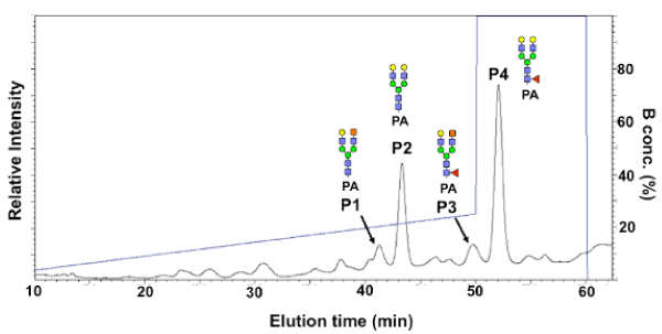
| Figure 5: HPLC analysis of PA-glycans. PA-glycans were separated by HPLC, and detected with a fluorescence detector (Ex: 320nm, Em: 400 nm). The elution pattern of reverse-phase HPLC with an Inertsil GL WP300 C18 column is indicated. The structures that were determined by MS are shown (P1 to P4). |