 |
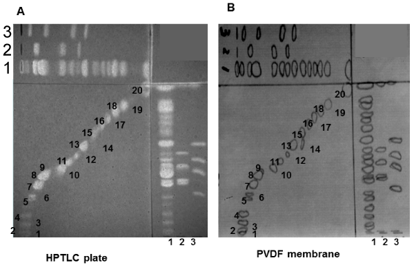
| Figure 1: The two-dimensional TLC-Blot of lipids from human hippocampus white matter. A, TLC of lipid from human hippocampus white matter, visualized with primuline. Lane 1,human hippocampus white matter; Lane 2, standard phospholipids, Car, PS, and SM from upper to lower band; Lane 3, standard phospholipids, PE, PI, PC, and LPC from upper to lower band. B, The visualized bands were marked with soft pencil and transferred to the PVDF membrane. |