|
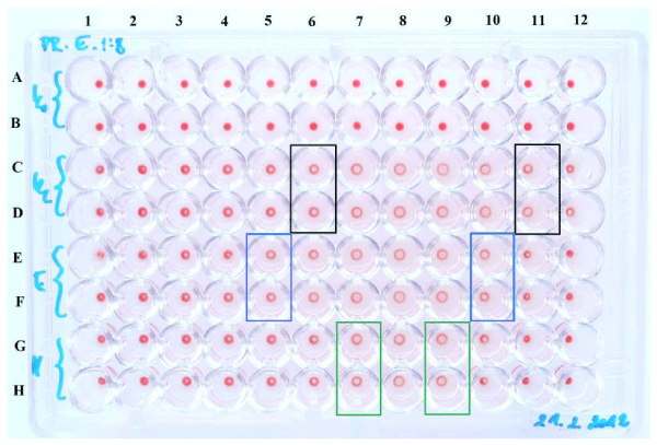
| Figure 2: Comparison of sialylation levels of erythrocytes. Lines A, B – negative control (no IBs); lines C, D – erythrocytes of Volunteer 1; lines E, F – erythrocytes of Volunteer 2; lines G, H – erythrocytes of Volunteer 3. Erythrocytes interact with 1,33-fold diluted IBs suspension (diluted in ratio 1:8) from column 1 to 12. Border wells of zones of positive hemagglutination are in frames, black frames – Volunteer 1; blue frame – Volunteer 2, green frame – Volunteer 3. |