|
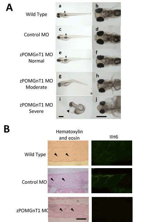
| Figure 2: Knockdown analysis of zPOMGnT1. (A) Embryos were injected with no sample (a, b), control MO (c, d), and zPOMGnT1 MO (e-j). In control MO-injected embryos, tail and eye developed normally (c, d). In zPOMGnT1 MO-injected embryos, normal phenotype showed normally developed tail and eye (e, f), moderate phenotype showed curved tail and small eye (g, h), and severe phenotype showed twisted tail, significant reduction in the size of eye and aberrant pericardium (i, j). Embryos were injected with each MO at the one- to two-cell stages and observed at 96 hpf. Arrowheads and arrow indicate swim bladder and pericardium, respectively. Scale bars = 500 μm. (B) Immunohistochemistry with anti-glycosylated α-DG antibody IIH6 in 96 hpf embryos. Left panels, hematoxylin and eosin staining; right panels, IIH6 staining. IIH6 immunoreactivity was detected in wild type and control morphant but decreased in zPOMGnT1 morphant. Arrowheads represent vertical myosepta. Scale bar = 50 μm. |