|
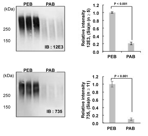
| Figure 2:Western-blotting analysis using polySia-specific antibodies. The PEB and PAB crude samples (10 μg as BSA/lane) were electrophoresed and blotted onto PVDF membrane. After blocking with 1% BSA, the membrane was probed with anti-polySia antibodies, 12E3 (1 μg/ml) specific to (Neu5Ac)5 or greater with non-reducing terminal end and 735 (0.8 μg/ml) specific to (Neu5Ac)11 were incubated. After washing, peroxidase conjugated anti-mouse IgG+IgM were incubated and color development was performed as described in Materials and Methods. Left panels show the results of Western-blotting. Right panels show the relative intensity of immunostained bands with antibodies and PEB value was set to 1.0. |