|
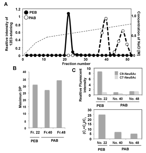
| Figure 5:Mono Q anion exchange chromatography of PEB and PAB. PEB and PAB were treated with monoSia-specific sialidase and subjected to the mono Q anion exchange chromatography. The samples were eluted with a NaCl gradient (0-10 min, 0 M; 10-30 min, 0 →0.3 M; 30-105 min, 0.3 →0.5 M; 105-135 min, 0.5 →3 M) in 20 mM Tris-HCl (pH 8.0). The flow rate was 0.5 ml/min and 1 ml was collected per fraction. After the samples were dot-blotted onto a PVDF membrane, the amount of polySia in each fraction was determined by the anti-polySia staining (12E3) of the membrane. (A) Chromatogram of mono Q anion exchange chromatography. (B) MH-FAEC analysis of peaks detected with 12E3 staining, fraction (fr.) 22 from PEB and fr. 40 and 48 from PAB. Maximum DP of the samples were shown. (C) Fluorometric C7/C9 analysis. Upper panel shows relative amounts of C7-Neu5Ac and C9-Neu5Ac. Samples derived from peaks detected with 12E3 staining, fraction 22 from PEB and fraction 40 and 48 from PAB were analyzed. Lower panel shows the (C9+C7)/C7 value, index as quality of di/oligo/polySia. |