|
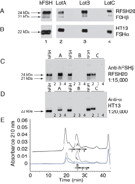
| Figure 3: Pituitary and urinary hFSH Western blots. Pooled human pituitary hFSH, commercially prepared postmenopausal gonadotropin, Pergonal, and individual first void urine characterized by Western blotting. A. Urinary hFSH lots analyzed with anti-hFSHβ antibody RFSH20. B. Urinary hFSH lots analyzed with anti-hFSHα antibody HT13. Lane 1, 1 μg hFSH (AFP4161B); lane 2, Pergonal lot A; lane 3, Pergonal lot B; lane 4, Pergonal lot C. C. RFSH20 Western blot of individual urinary hFSH preparations shown in panel E, below. D. HT13 Western blots of urinary hFSH, as above. Column fractions as indicated below. E. Superdex 75 chromatograms showing purification of anti-hFSHβ 46.3H6.B7-bound hFSH from first void urine samples. The bars show portions of the chromatograms analyzed by Western blotting in panels C and D, above. |