 |
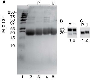
| Figure 4: SDS-PAGE and Western blot of pituitary and urinary hFSH preparations. Samples consisting of 1 or 10 μg hFSH were reduced in the presence of 5% β-mercaptoethanol and 1% SDS, and subjected to electrophoresis on 15% polyacrylamide gels. A. Proteins visualized by Coomassie blue staining. Based on these results, the pituitary hFSH preparation AFP7298A was selected for glycan analysis. Lane 1, Bio-Rad Precision Plus high MW pre-stained MW markers; lane 2, 10 μg pituitary hFSH (AFP7220); lane 3, 10 μg pituitary hFSH (AFP7298A); lane 4, 10 μg pituitary hFSH (AFP5720); lane 5, 10 μg urinary hFSH. B. FSHβ subunits visualized by Western blotting using monoclonal antibody RFSH20, diluted 1:10,000. C. FSHa subunit Western blot using monoclonal antibody HT13. Lane 1, 1 μg pituitary hFSH (AFP7298A); lane 2, urinary hFSH. While all images were acquired with a BioRad VersaDoc 4000, the positions in the instrument and lenses employed for transillumination and chemiluminescence imaging were different, thereby producing different sized images. |