|
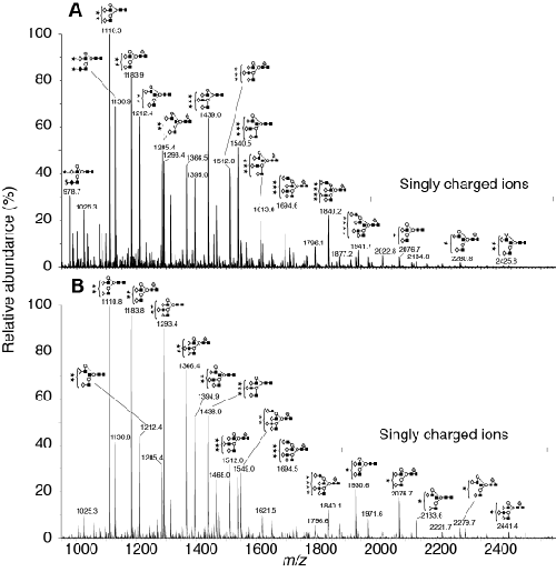
| Figure 5: ESI-mass spectrometry analysis of intact pituitary and urinary hFSH glycans. A. Negative ion ESI spectrum of pituitary hFSH glycans. B. Negative ion ESI spectrum of urinary hFSH oligosaccharides. Neutral and acidic glycan masses along with compositions are listed in Tables 1 and 2. |