|
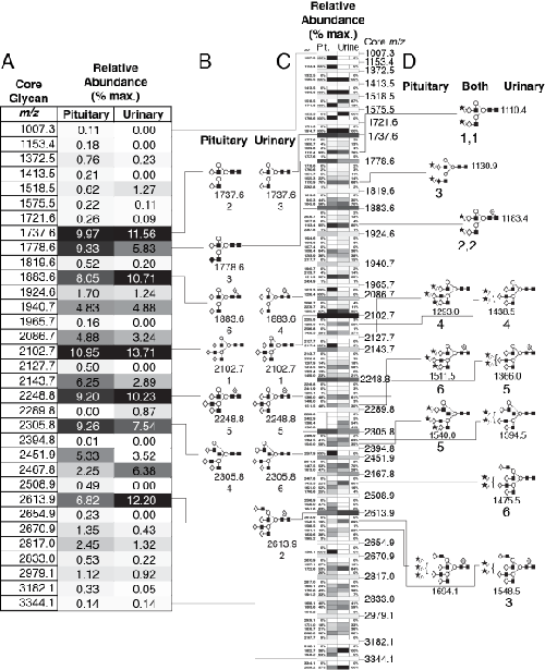
| Figure 7: Comparison of pituitary and urinary hFSH glycan populations. A. Relative abundance of glycans based on glycan core structure indicated by gray scale. Each bar represents the relative abundance of the sum of glycan variants possessing the same underlying glycan structure for a given hormone (Table 1) both as shaded (0 = white, 100% of max. = black) and numerical values. B. Core glycan structures for the 6 most abundant glycan families in panel A. The m/z value and rank order of relative abundance are given below each. C. Glycan variant abundance within each glycan family (wide bars). Narrow bars indicate relative abundance of each variant using gray scale 0-100% and shown in the same order listed in Table 1. The glycan variant m/z and relative abundance within each group are indicated beside each bar and are intended for digital format, which permits zooming in to read individual values. The wide bars represent each glycan family listed in panel A, and are identified by the m/z values on the right. These serve to separate each group of individual glycan variants. D. Glycan structures for the 6 most abundant glycan variants in each FSH preparation. When the same variant is most abundant in both pituitary and urinary hFSH, the structures are centered. When they differ, pituitary glycans are shown on the left and urinary glycans on the right. The numbers indicate the order of abundance within each FSH preparation. Families 3-5 and variants 3-5 were very similar in abundance. |