 |
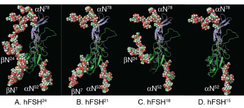
| Figure 2: Human FSH glycoform models. The FSHα (green) and FSHβ (blue) subunits are shown as backbone cartoons. The N-glycans are shown as spheres and represent the most abundant glycans observed in glycopeptide mass spectra [6]. Panel A. hFSH24, which possesses all 4 N-glycans. Panel B. hFSH21, which lacks βAsn24 glycan. Panel C. hFSH18, which lacks βAsn7 glycan. Panel D. hFSH15, which lacks both FSHβ N-glycans. The hFSH24 model was created using Tripos Sybyl and subjected to molecular dynamics. The image in panel A was rendered with PyMol and the FSHβ glycans hidden in subsequent panels. |