 |
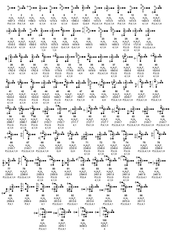
| Figure 4: Diagrams showing the structures of the neutral core glycans associated with the 6 hFSH and hFSH glycoform preparations analyzed in this study. The m/z value for the ion associated with the neutral glycan and composition are shown with each structure. In several cases, more than one structure was possible and either could not be eliminated by MS/MS analysis or else all were shown to be present by MS/MS characterization. FSH preparations in which at least 1 glycan ion was detected that shared the same core glycan structure are indicated as follows: P = pituitary FSH, U = urinary FSH, G = recombinant GH3-hFSH, 4 = hFSH24, 1 = FSH21, and H = hFSH21/18 from hLH preparations. |