 |
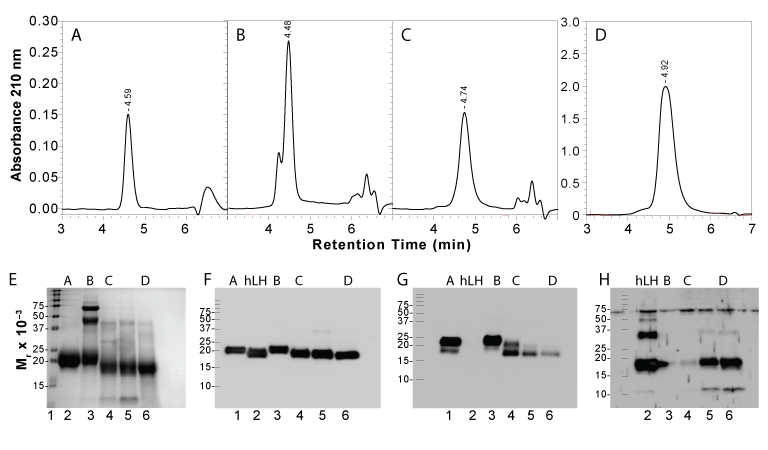
| Figure 10: Size exclusion chromatography and Western blot characterization of hFSH. A-D. Samples of hFSH were subjected to UPLC-SEC using a 0.46 x 15 cm Waters BEH200 SEC column equilibrated with 20% acetonitrile in 0.2 M ammonium bicarbonate at a flow rate of 0.4 ml/min. A. 0.5 μg GonalF recombinant hFSH; B. 1.5 μg pituitary hFSH24; C. 1 μg pituitary hFSH21; D, 20 μg pituitary hFSH21. E-H. Electrophoretic characterization. E. SDS-PAGE with Coomassie Blue staining. Lane 1, BioRad broad MW pre-stained standards; 2, 10 μg pituitary hFSH AFP-7298A; lane 3, 10 μg pituitary hFSH24 from panel B; lane 4, 10 μg hFSH24 from panel C; lane 5, 10 μg pituitary hFSH21; lane 6, 10 μg pituitary hFSH21 from panel D. F. Common -subunit Western blot probed with monoclonal antibody HT13 diluted 1:4000. G. FSH? Western blot probed with monoclonal antibody RFSH20 diluted 1:10,000. H. LH? Western blot probed with anti-eLH? antibody VB-I-118 diluted 1:1000. Lane 1, 1 μg pituitary hFSH AFP-7298A (missing in panel H); lane 2, 1 μg hLH; lane 3, 1 μg pituitary hFSH24 from panel B; lane 4, 1 μg pituitary hFSH21 from panel C; lane 5,1 μg pituitary hFSH21 ; lane 6, 1 μg pituitary hFSH21 from panel D. |