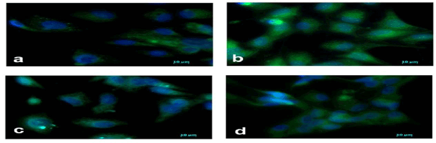
a: control (untreated), b: AGE (100 μg/ml) treated c : Glycine (2.5 mM)+AGE (100 μg/ml),d: Glutamic acid (2.5 mM)+AGE (100 μg/ml).
The green fluorescence represents the phalloidin stained active actin stress fibers. Nucleus is stained blue with DAPI
The Stress fiber formation showed an increase with AGE compared to control. Glycine and glutamic acid decreased the actin stress fiber formation when added along with AGE (100 μg/ml).