 |
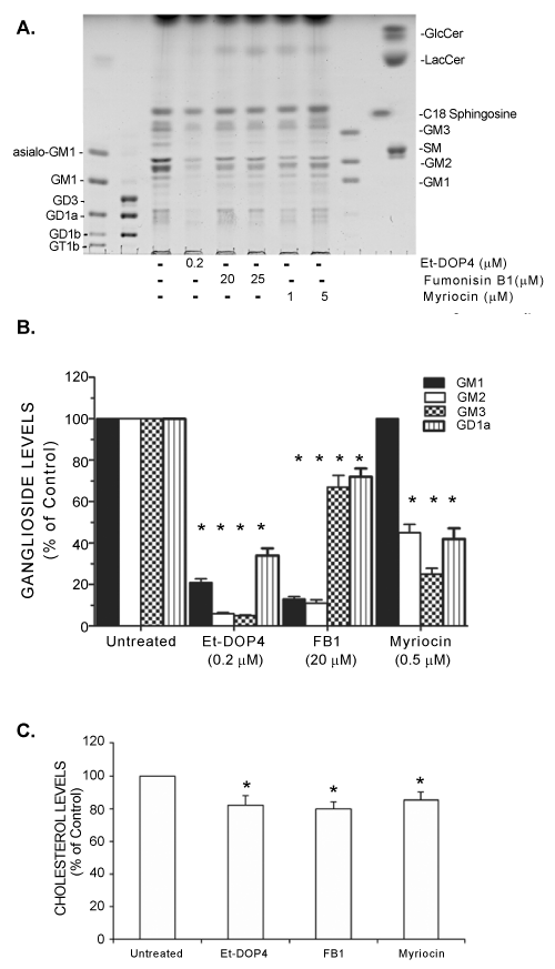
| Figure 2: Depletion of gangliosides from cultured ECV304 cells with sphingolipids synthesis inhibitors: The lipid sample preparation and thin layer chromatographic analysis were detailed in the Methods section. A. A representative high performance thin layer chromatography plate displaying the ganglioside pattern of cultured EV304 cells after a 48-h treatment with inhibitors. B. Comparison of ganglioside levels in untreated and treated ECV304 cells with sphingolipid inhibitors. The data represent the mean ± SEM from 3 separate experiments. * denotes p< 0.05 as determined by twoway ANOVA using GraphPad Prism. C. The reduction of cholesterol content in inhibitor treated ECV304 cells. Crude cellular lipids were normalized to 20 nmol phospholipid phosphate prior to separation by high performance thin layer chromatography using a solvent system consisting of chloroform/acetic acid (9:1, v/v). The plates were scanned and analyzed ImageJ software, and the levels of cholesterol were quantified by comparison tountreated control samples. |