|
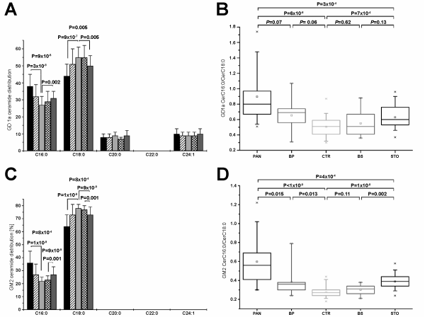
| Figure 4: Ceramide profiles and CerC16:0/CerC18:0 ratios of serum gangliosides GD1a and GM2. Ceramide signals are normalized based on the sum of all structures detected among (A) GD1a and (C) GM2 for each sample. CerC16:0/CerC18:0 ratios of (B) GD1a and (D) GM2 of individual patients are displayed as boxplots. Patient groups are represented as follows: pancreatic cancer (black/PAN), benign pancreatic diseases (striped/BP), non-diseased controls (white/CTR), benign gastric diseases (checked/BS), gastric cancer (grey/STO). Error bars indicate standard deviations. P-values displayed in the upper panel of each diagram (panels A and C only for P≤0.01) were obtained by the non-parametric Mann-Whitney-U test. |