 |
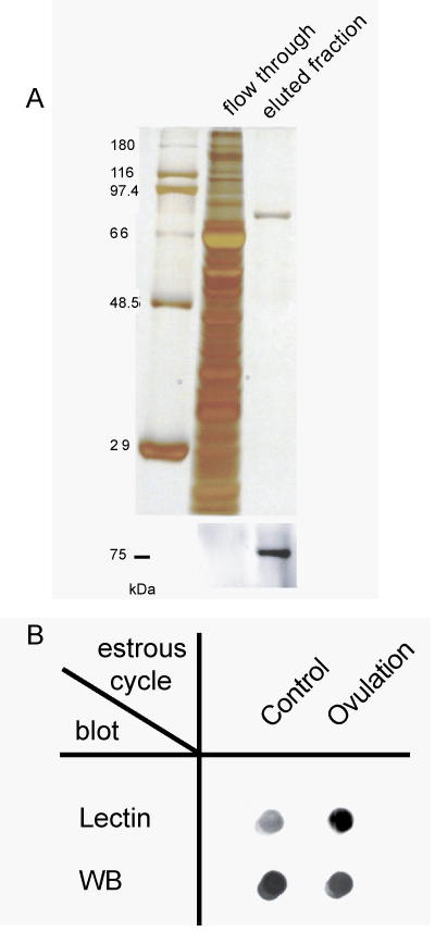
| Figure 4: Comparison of sialylation of transferrin. (A) After applying the modified protocol, transferrin was recovered from the eluted fraction by the immunoprecipitation method. Upper is silver staining. Lower is western blot analysis using an anti-transferrin antibody. (B) Lectin blot analysis using Sambucus sieboldiana agglutinin was performed on ovaries to check the total sialic acid quantity. Western blot analysis was performed using the antitransferrin antibody. |