 |
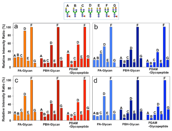
| Figure 4: Comparative analysis of abundance ratios of glycan structures among three kinds of derivatization detected by MS. a, the signal pattern of glycans A-G in positive-MALDI-QIT-TOF MS. The structures of glycans A-G are indicated on the top. The relative values are shown when glycan F signal is 100%. b, the signal pattern of glycans A-G in negative-MALDI-QIT-TOF MS. c, the signal pattern of glycans A-G in positive-MALDI-TOF MS in linear mode. d, the signal pattern of glycans A-G in negative-MALDI-TOF MS in linear mode. |