 |
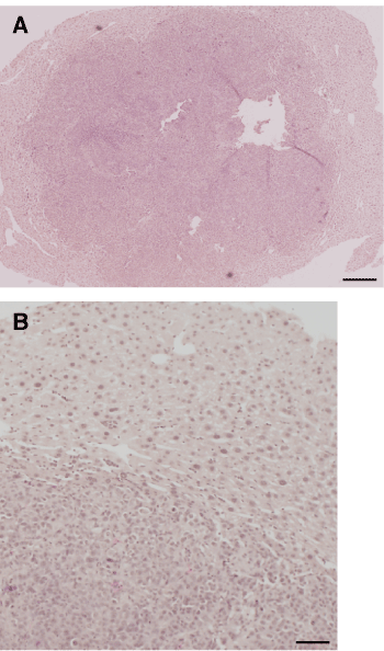
| Figure 2: HE staining of Hepa 1-6 liver tumor. (A) For liver tumor formation, Hepa 1-6 cells were suspended in DMEM and injected under the capsule of the spleen in three 9-week-old BALB/c nu/nu mice. After 4 weeks, metastatic liver tumors were removed, fixed in 4% paraformaldehyde, and stained with HE. Scale bar, 250 μm (B) High-power field. Scale bar, 100 μm. |