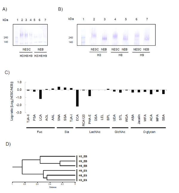
A) The soluble extracts from the three hESCs (H3, H8 and H9) and the corresponding hEBs were directly eluted on lanes 2-4 and 5-7, respectively, and were subjected to western blot analysis using anti-podocalyxin pAb.
B) Immunoprecipitated podocalyxin from hESCs and hEBs were subjected to western blot analysis with anti-podocalyxin pAb. The precipitates from three hESC lines were eluted on lanes 2 (H3), 4 (H8) and 6 (H9), and those from the corresponding hEBs were eluted on lanes 3 (H3), 5 (H8) and 7 (H9).
C) Lectin microarray data were max-normalized, and the log indexes of individual lectins are compared as in figure 3A.
D) Hierarchical clustering analysis showing two clusters of hESCs and hEBs.