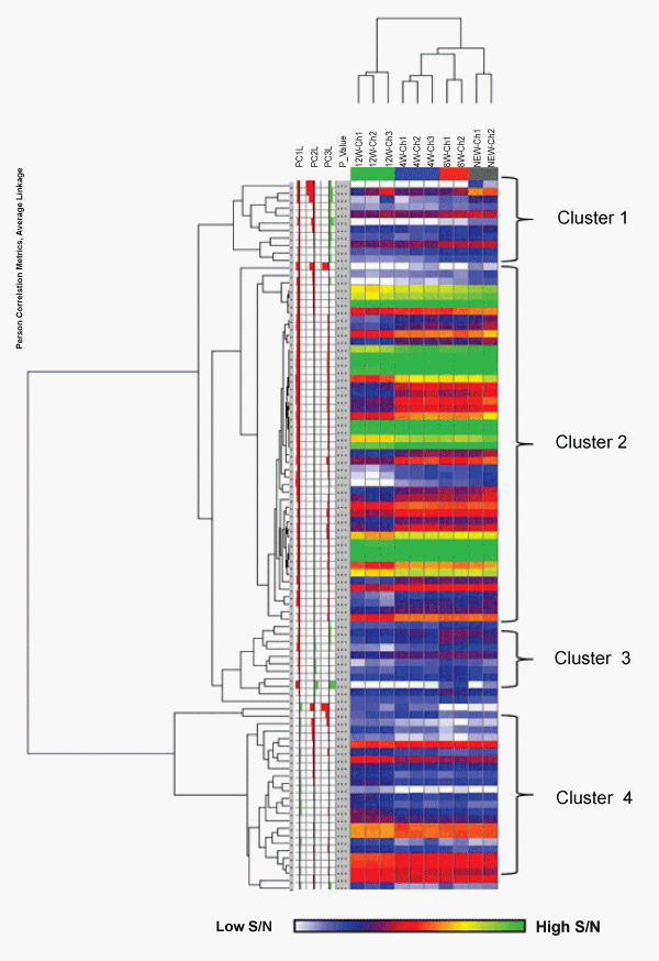
This HCA analysis was based on 95 masses showing significant differences with p value < 0.01. Cells are colored according to the signal to noise (S/N) intensity (log2 scaled). Masses clustered using a Pearson correlation, samples clustered using Euclidean correlation. Light blue - low intensity, dark blue/purple/red - medium intensity, orange/yellow/green - highest intensity.