 |
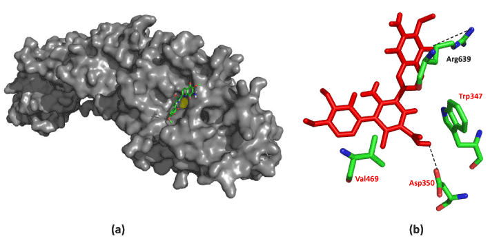
| Figure 6: Binding conformation of streptonigrin inside the PAD4 binding pocket. Streptonigrin is seen occupying the binding pocket with Cys645 in yellow (a). PAD4 residues with hydrophobic contacts with streptonigrin (red, stick structure) are labeled in red, while hydrogen bonds are drawn as dashed line (b). |