|
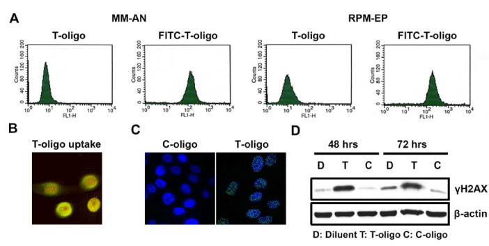
| Figure 1: Uptake of T-oligo and its effects on MM-AN melanoma cells. MM-AN cells were treated with either diluent or 50 μM of FITC labeled T-oligo for 4hours and uptake of T-oligo was studied. A. FACS analysis demonstrated the uptake of T-oligo by MM-AN cells and RPM–EP cells. B. Confocal microscopy of MM-AN cells showing nuclear localization of FITC-T-oligo. The nucleus was stained with propidium iodide and colocalization of the two fluorescent molecules was seen as a yellow color. C. MM-AN melanoma cells were treated with T-oligo or c-oligo for 48 hours, fixed in paraformaldehyde, and immunostaining was performed with a ?H2AX monoclonal antibody and a secondary antibody linked with FITC. ?H2AX foci were seen mainly in cells treated with T-oligo. Very few foci could be detected in cells treated with c-oligo.D. MM-AN melanoma cells were treated with T-oligo, c-oligo, or an equal volume of diluent for the indicated times and samples were collected for western blotting for detecting ?H2AX (Phospho Ser-139 H2AX). ?H2AX was upregulated at 48-72 hours after treatment with T-oligo. |