 |
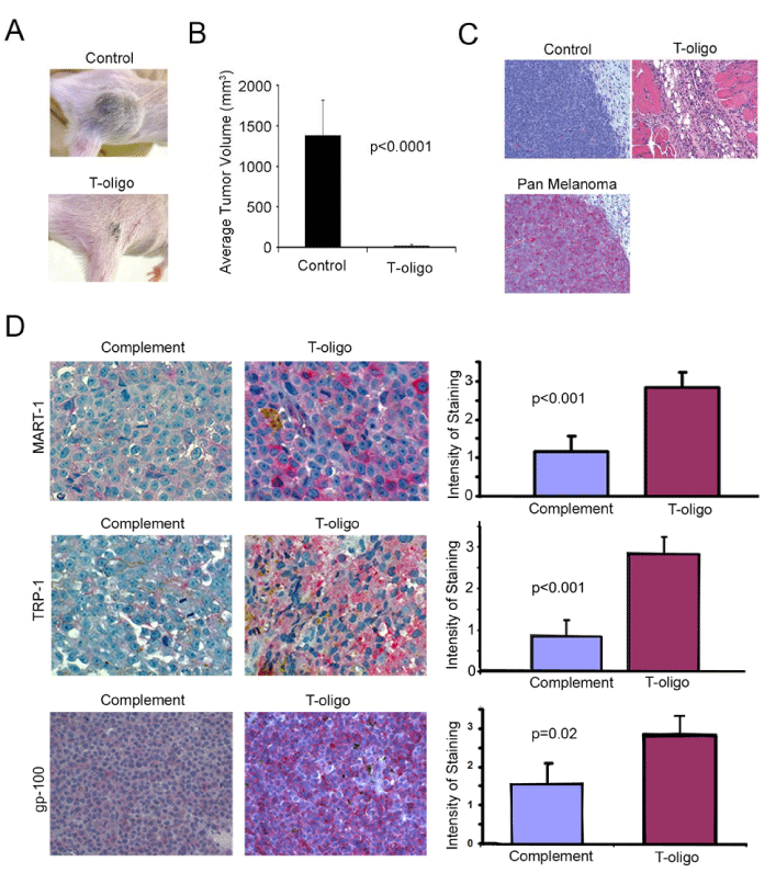
| Figure 4: Reduction of tumor size and upregulation of differentiation markers with T-oligo treatment: One week after injection of MM-AN cells, SCID mice with visible melanoma tumors on the flank were treated with 420 μg of T-oligo or c- oligo daily for one week and then left untreated for two weeks. A. T-oligo treated mice showed very small residual tumors in comparison to animals treated with c-oligo. B. Animals (n=10) treated with T-oligo showed a 98% reduction in tumor volume as compared to c-oligo. The differences in tumor size in these studies was statistically significant (p<0.0001) C. H and E staining of animals treated with T-oligo and c-oligo showed that animals treated with c-oligo had large tumors. Tumors were absent in 60% of animals treated with T-oligo. Melanoma tumors were identified by their positive staining with the pan melanoma cocktail. D. Sections from FFPE tumors from SCID mice were prepared and immunostaining procedures were performed. FFPE sections incubated with non-immune rabbit serum served as negative controls. T-oligo treatment increased expression of MART-1, TRP-1 and gp-100. |