|
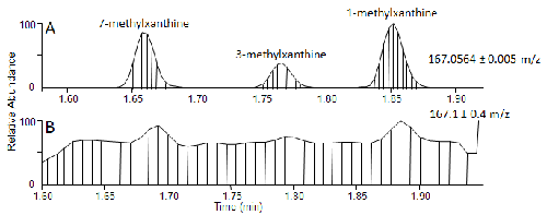
| Figure 3: ESI+ extracted ion chromatogram of the mass for the methylxanthine family of isomers from a human serum extract from the HRAM instrument (A) and from the UMR instrument (B). Precise mass and mass windows used are labeled on each panel. The added sensitivity provided by increased mass resolution and accuracy permitted the detection of all three methylxanthine isomers in the HRAM data streams from the inherent background noise, which was not possible with the UMR data. Vertical lines indicate full MS scans taken during analysis. Note the faster scan speed of the HRAM instrument. |