|
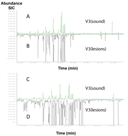
| Figure 3: Inversely overlaid selected ion m/z 55 chromatograms at C-F (sound) and C-A (lesions) sites of two subjects after glucose solution rinse. Y-axis shows the abundance (intensity of the signal) and x-axis shows the elapsed time. A, B: subject 1; C, D: subject 2. |