|
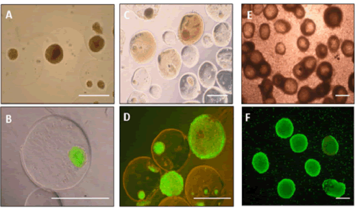
| Figure 3: Representative images of fresh and retrieved microencapsulated islets Light microscopic (A,C,E) and fluorescent microscopic images (B,D,F) of microencapsulated islets. Pictures were taken after encapsulation (A,B), at 76 days after transplantation into BALB/c mice (C,D), or at 20 days after transplantation into NOD mice (E,F). Viability staining with fluorescein diacetate (FDA; green=viable) and propidium iodide (PI; red=dead) of encapsulated islets. Bar scale- 500μm. |