|
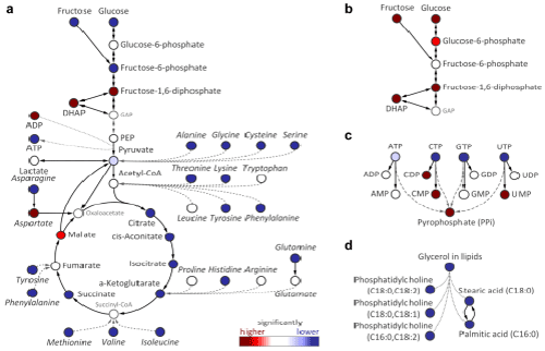
| Figure 2: ANOVA comparison of different sampling methods across both cell lines. For a given comparison between two sampling methods red metabolite nodes signify higher metabolite levels while blue nodes signify lower levels with a significance of q values <0.1 for least intensive colors to q value <0.01 for most intensive colors.Grey metabolites were not measured and grey dashed arrows implicate several enzymatic reaction steps which were omitted for the sake of clarity. A) Comparison of trypsinization versus MxP® CellCollect reveals differences in glycolysis, TCA-cycle and protein genic amino acids. B), C), D) Comparison of scraping versus MxP® CellCollect with B) focusing on first steps of glycolysis, C) summarizing results for nucleoside tri-, di- and monophosphates and d showing results of selected lipid metabolites and their fatty acid precursors. The glycerol in lipids describes the sum of all glycerol moieties that is released from the lipid fraction of a biological sample after hydrolysis of complex lipids and subsequent derivatization prior to the gas chromatography analysis. Several phosphatidylcholines are shown as examples together with two saturated fatty acids which represent the sum of its occurrence both in free and in all lipid-bound forms (dotted lines indicate that fatty acids and phosphatidylcholines contribute to the total sum of conjugated glycerol in complex lipids). |